The 0.1% of Your Brain That Decides You Need Sleep
A tiny population of cortical neurons—one in a thousand—can override wakefulness and initiate deep sleep from inside the cortex itself.
By The Breakthroughist Science Desk
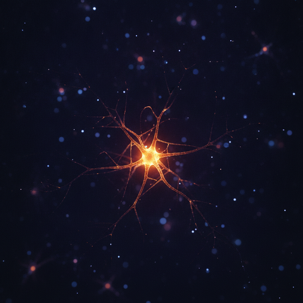
Renata Batista-Brito had been chasing a cell type so rare that grant applications kept getting rejected for being too high-risk. Sst-Chodl neurons make up roughly one neuron in a thousand in the cerebral cortex [1]. Her lab spent years building the molecular strategy to find them. When she finally looked at the anatomy, she expected to see very little. Instead, she saw two or three cell bodies in the whole brain—and branching so dense and far-reaching she had no reference point for it. "At first, I saw two or three cell bodies in the whole brain," Batista-Brito told Ars. "Despite that, there was massive, massive arborization all over the visual cortex, like I never saw with any other neuron." [1]
The implications of that anatomy now extend well beyond cell biology. "What our work shows is that the cortex can not only see this rhythm but also initiate it by itself, and this is sufficient to promote sleep," Terral said [1].
Sleep science has long drawn a clean organisational chart, with the cortex near the bottom. The prevailing assumption was that the signals controlling sleep descended from deeper structures, and the cortex simply received them. What happens when a cell type hiding in plain sight rewrites that hierarchy from the inside?
For decades, sleep research treated the cerebral cortex as a passive follower reacting to signals from the deep brain. "Usually, sleep is associated with being controlled by subcortical regions," Terral said [1]. The cortex is where the slow rhythms of deep sleep can be seen in electrical recordings—but researchers assumed the signals that triggered them originated elsewhere, in structures like the hypothalamus and the thalamus. The Sst-Chodl finding challenges that assumption directly [1].
The neurons get their name from two genes—Sst and Chodl—that are simultaneously active in them [1]. Long-range inhibitory neurons carrying these genetic signatures had been known for some time, based on studies in monkeys, but researchers couldn't figure out how to manipulate them using these genes. The problem was specificity. "A single gene is not able to target these cells," Batista-Brito said [1]. Inhibitory neurons already account for only about a fifth of all cortical neurons, so Sst-Chodl cells work out to roughly one neuron in a thousand in the cortex [1]. Any contamination in the targeting strategy would drown the signal. "If you're not really specific, the contaminants are going to be much more dominant than the specific cells," Batista-Brito said [1]. Building a labelling strategy that activates only when both genes are expressed simultaneously took her team years [1].
When the team finally imaged the cells' anatomy, what they found defied the standard picture of how inhibitory neurons are wired. Most inhibitory neurons are local: they receive signals from sometimes-distant regions and exert control over their immediate surroundings—a small patch of nearby tissue [1]. Sst-Chodl neurons work in the opposite direction. "They are receiving inputs that are quite precise, but then they are broadcasting that information everywhere," Batista-Brito said [1]. A single Sst-Chodl cell branches out across the entire visual cortex, then sends axons to areas responsible for touch, hearing, spatial memory, navigation, and voluntary movements [1].
A neuron that listens narrowly and speaks broadly is an unusual piece of biological architecture. It suggests a coordinating function—less a local gatekeeper and more a signal that tells an entire distributed system to change its mode of operation simultaneously.
To map when the cells are active, the team imaged them while simultaneously tracking pupil size, muscle tone, facial movement, running, and cortical electrical activity in mice [1]. Ninety-five out of 111 imaged cells lit up during slow-wave sleep and quiet, motionless wakefulness, and fell silent during running and REM sleep [1]. The firing pattern during slow-wave sleep was especially striking. During that state, the cortex alternates between UP states—periods of vigorous neural firing—and DOWN states of near-silence. Sst-Chodl neurons broke from that pattern. "Their activity is even higher at the termination of the UP state," Terral said. "Once all the other neurons start to decrease their activity, those neurons start to increase it even more." [1]
The team then used optogenetics—inserting a gene for a light-activated ion channel so that neurons can be made to fire on command—to test what Sst-Chodl stimulation actually does to the brain [1]. The results were precise in a way that simple rate-based models of neural control would not predict. Stimulating the cells drove delta power—the slowest, highest-amplitude brain waves associated with deep, restorative sleep—up across every cortical layer [1]. But overall firing rates barely changed. "Those neurons are not changing so much the firing rate," Terral said. "They change just the coordination—whether the neurons fire together or not." [1]
That distinction between rate and coordination matters. Normally, when researchers drive a neural population at a given frequency, the surrounding network entrains to that frequency. Here it didn't. "If we did a flat stimulus, or delta, or 20 hertz, or 60 hertz, you still induced the same kind of oscillation," Batista-Brito said [1]. The cells appear to have intrinsic properties that lock them to a specific output regardless of input frequency—though the team is candid that the mechanism is still unknown [1]. The reach of their influence also exceeded what anatomy alone would suggest. Direct inhibitory currents appeared in a third of cells two millimeters out, and effects were measurable as far away as frontal motor areas [1].
The behavioural experiments were the most direct test of the cells' function. Activating Sst-Chodl neurons across the cortex of freely moving mice increased both slow-wave and REM sleep, cut the time taken to fall asleep, and sent the animals into their nests during the day [1]. Then the team ran the same experiment during the dark phase, when nocturnal mice are predominantly awake. "We could have them sleep more during the time that they are awake than they usually sleep during the day," Batista-Brito said [1].
Sleep, Batista-Brito explains, comes in two distinct forms: the circadian variety tied to light cycles, and homeostatic sleep driven by how long a person or animal has been awake [1]. Kilduff showed that after sleep deprivation, Sst-Chodl cells are the most active neurons in the cortex [1]. That finding positions these cells not just as sleep initiators but potentially as the biological mechanism by which the brain monitors how badly it needs rest.
The paper's scope comes with acknowledged limits. All the work was carried out in the visual cortex, and while the team expects the coordinating role of Sst-Chodl neurons to generalise across brain regions, the precise wiring may differ [1]. Batista-Brito's lab is now repeating the anatomical work in the prefrontal cortex. Their prediction is that prefrontal Sst-Chodl cells receive inputs from the hypothalamus and the thalamus—and that those inputs are what specifically triggers the sleep network in that region [1]. Three core questions remain open: what activates these cells in the first place, whether they genuinely sense sleep pressure, and why they drive delta power [1].
Open questions are, in their own way, the most useful output of a study like this. A finding that closes a gap cleanly is satisfying; a finding that redraws the entire map of where to look next is something more durable. The cortex has just acquired a much more interesting interior life.
The broader clinical stakes are considerable. Sst-Chodl neurons are conserved across species, from salamanders to humans, which the paper argues makes them a plausible entry point into the sleep disruption that runs through so many psychiatric illnesses [1]. The team published their results in Nature, DOI: 10.1038/s41586-026-10876-y [1]. If Batista-Brito's lab can confirm that these cells behave similarly in the prefrontal cortex—and determine what activates them—the path toward understanding, and potentially addressing, disordered sleep opens at an entirely new level of biological precision.
"Going after these cells was really a high-risk project because the likelihood of seeing anything with 0.1 percent of neurons in the cortex is really low," Batista-Brito said [1]. "I wrote a bunch of grants on these projects that were always rejected because it was too high-risk." The rejected proposals look different now that one neuron in a thousand has been shown to carry the cortex's instruction to sleep.
They are receiving inputs that are quite precise, but then they are broadcasting that information everywhere.Renata Batista-Brito